Skip to content
  • Our Work
    • Fields
      • Cardiology
      • ENT
      • Gastro
      • Orthopedics
      • Ophthalmology
      • Pulmonology
      • Surgical
      • Urology
      • Other
    • Modalities
      • Endoscopy
      • Medical Segmentation
      • Microscopy
      • Ultrasound
  • Success Stories
  • Insights
    • Magazine
    • Upcoming Events
    • Webinars
    • Meetups
    • News
    • Blog
  • The company
    • About us
    • Careers
Menu
  • Our Work
    • Fields
      • Cardiology
      • ENT
      • Gastro
      • Orthopedics
      • Ophthalmology
      • Pulmonology
      • Surgical
      • Urology
      • Other
    • Modalities
      • Endoscopy
      • Medical Segmentation
      • Microscopy
      • Ultrasound
  • Success Stories
  • Insights
    • Magazine
    • Upcoming Events
    • Webinars
    • Meetups
    • News
    • Blog
  • The company
    • About us
    • Careers
Contact

Middle and Inner Ear Segmentation with Deep Learning

Ear pathologies are common in all age groups, and are one of the leading causes for visiting a doctor. In most cases, proper diagnosis can be achieved via clinical examination. However, in recurrent or complicated cases imaging of the ear is indicated and can help with treatment choice.


Inner ear segmentation
Inner Ear segmentation: semicircular canals (red) and cochlea (green)

The human ear is in charge of two separate functions – hearing and maintaining balance. Its complex structure includes the outer ear which collects sound waves, middle ear which conducts the sound to the inner ear, that is located within the temporal bone, and consists of the vestibular apparatus and the cochlea.

Due to its intricate structure, many pathologies can potentially arise within the different parts of the ear, for which imaging such as computed tomography (CT) or magnetic resonance imaging (MRI) may be indicated. CT aides with assessing the osseous structures of the ear, while MRI is especially good for imaging of the vestibulocochlear nerve, the brain and other fluid-filled spaces. Ear imaging is useful for evaluation of congenital sensorineural hearing loss, vestibular schwannoma and cholesteatoma.

Imaging is also used for surgical planning by allowing the surgeon to better visualize the tumor that needs to be resected, or choosing the best drilling trajectory and appropriate electrode measurements for cochlear implant surgery. Moreover, 3D segmentation of the middle and inner ear may help with minimizing surgical complications including damage to nearby vessels or nerves. For example, damage to the facial nerve could result in facial paralysis.

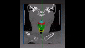
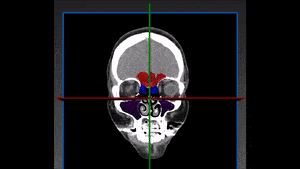
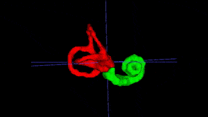
Therefore, 3D segmentation of the ear generated from CT and MRI images is useful for demonstrating the complex anatomical structures, and by doing so – minimizing complications and allowing better surgical planning and precise diagnosis.

Automated Middle and Inner Ear Segmentation

For this task, a proprietary deep learning model based on a fully convolutional neural network was developed by RSIP Vision. The complexity of the structures and their fine details are met by the network thanks to the cutting edge technology implemented within our model. Take us along for you next deep learning project!

Share

Share on linkedin
Share on twitter
Share on facebook

Main Field

ENT

View ENT

Categories

  • ENT, RSIP Vision Learns

Related Content

Larynx

Larynx Segmentation with Deep Learning

Sinus segmentation

Sinus Segmentation with Deep Learning

Larynx

Larynx Segmentation with Deep Learning

Sinus segmentation

Sinus Segmentation with Deep Learning

Show all

RSIP Vision

Field-tested software solutions and custom R&D, to power your next medical products with innovative AI and image analysis capabilities.

Read more about us

Get in touch

Please fill the following form and our experts will be happy to reply to you soon

Recent News

IBD Scoring – Clario, GI Reviewers and RSIP Vision Team Up

RSIP Neph Announces a Revolutionary Intra-op Solution for Partial Nephrectomy Surgeries

Announcement – RSIP Vision Presents Successful Preliminary Results from Clinical Study of 2D-to-3D Knee Bones Reconstruction

Announcement – New Urological AI Tool for 3D Reconstruction of the Ureter

All news
Upcoming Events
Stay informed for our next events
Subscribe to Our Magazines

Subscribe now and receive the Computer Vision News Magazine every month to your mailbox

 
Subscribe for free
Follow us
Linkedin Twitter Facebook Youtube

contact@rsipvision.com

Terms of Use

Privacy Policy

© All rights reserved to RSIP Vision 2023

Created by Shmulik

  • Our Work
    • title-1
      • Ophthalmology
      • Uncategorized
      • Ophthalmology
      • Pulmonology
      • Cardiology
      • Orthopedics
    • Title-2
      • Orthopedics
  • Success Stories
  • Insights
  • The company