Skip to content
  • Our Work
    • Fields
      • Cardiology
      • ENT
      • Gastro
      • Orthopedics
      • Ophthalmology
      • Pulmonology
      • Surgical
      • Urology
      • Other
    • Modalities
      • Endoscopy
      • Medical Segmentation
      • Microscopy
      • Ultrasound
  • Success Stories
  • Insights
    • Magazine
    • Upcoming Events
    • Webinars
    • Meetups
    • News
    • Blog
  • The company
    • About us
    • Careers
Menu
  • Our Work
    • Fields
      • Cardiology
      • ENT
      • Gastro
      • Orthopedics
      • Ophthalmology
      • Pulmonology
      • Surgical
      • Urology
      • Other
    • Modalities
      • Endoscopy
      • Medical Segmentation
      • Microscopy
      • Ultrasound
  • Success Stories
  • Insights
    • Magazine
    • Upcoming Events
    • Webinars
    • Meetups
    • News
    • Blog
  • The company
    • About us
    • Careers
Contact

Cardiovascular Ultrasound Software

Heart failure is the leading cause of mortality in the western world. Routine examination in clinics can greatly reduce the risk of heart failure by recognizing early symptoms, correcting classification and assigning appropriate treatment. Monitoring the heart during a clinic visit can shed light onto the causes and symptoms leading to heart disease. Monitoring can be performed by noninvasive and relatively short operation using electrophysiological sensors (ECG) and echocardiogram (e.g. ultrasound). Problems stemming from pulse irregularities (e.g. arrhythmia) can be analyzed using electrocardiograms; however, a visual examination teaches the examining physician a great deal about the continuous operation of the heart during operation and malfunction.

Cardiovascular Ultrasound software at work

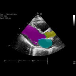
The volume of the heart’s ventricles at the end diastole and systole serve as prognostic indicators for arrhythmia, ventricular fibrillation and the risk for cardiac arrest. Extracting information regarding the dynamics of ventricles contraction is therefore important for the diagnosis of these pathologies. Ultrasound technology offers a straightforward view of ventricles dynamics, so that cardiovascular ultrasound devices are common in clinical practice for initial examination and quick prognosis. The image obtained from ultrasound is generated by collecting the reflected sound waves off of tissues into an intensity map. Advances in image ultrasound reconstruction allows better visualization of tissues using nondestructive frequencies; however ultrasound images remain noisy and their interpretation requires an experienced physician.

.

Automated cardiovascular ultrasound software

RSIP Vision has constructed an automated cardiovascular ultrasound procedure to track the ventricles throughout the heart’s cycle. Ultrasound recording in a 4-chamber view over a period of several heart cycles allows RSIP Vision’s algorithm to track the contraction and report frequencies and volume, thus serving as a quantitative tool to assist in delivering accurate prognosis.

Tracking the edges of the left and right ventricles in a noisy ultrasound is not a trivial task. In addition to the noise, the deformable nature of the heart, the translation caused by breathing and the operator moving the ultrasound device, all add to the complication. RSIP Vision’s cardiovascular ultrasound software compensates for these types of involuntary motion and successfully tracks the ventricle walls.

.

Ultrasound software advanced techniques

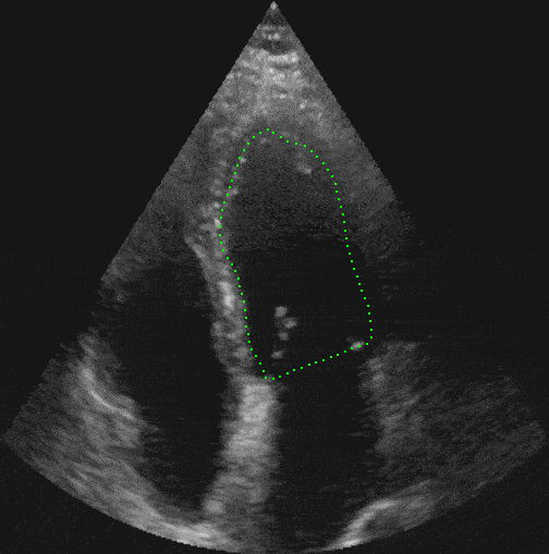
The technique we chose to track deformable objects is active contour models (snakes). The active contours tend to cling on the ventricle walls during contraction by minimizing a tailor made energy functional, which takes its image specific gradient and intensity maps as input. Successful tracking allows us to segment the ventricles and therefore report on the dynamics of the volume as it changes throughout the heart cycle. Embedding our algorithm in an ultrasound station, quantitative measure can be obtained in real-time.

RSIP Vision has been developing customized image processing and computer vision software and solutions for cardiology and other medical fields for over 2 decades. You are invited to contact our engineering team to learn how RSIP Vision can help you achieve your R&D goals.

Share

Share on linkedin
Share on twitter
Share on facebook

Main Field

Ultrasound

View Ultrasound

Categories

  • Cardiology, RSIP Vision Learns, Ultrasound

Related Content

Deep Learning for Cardiac Ultrasound (Echocardiography)

Deep Learning for Cardiac Ultrasound (Echocardiography)

Show all

RSIP Vision

Field-tested software solutions and custom R&D, to power your next medical products with innovative AI and image analysis capabilities.

Read more about us

Get in touch

Please fill the following form and our experts will be happy to reply to you soon

Recent News

IBD Scoring – Clario, GI Reviewers and RSIP Vision Team Up

RSIP Neph Announces a Revolutionary Intra-op Solution for Partial Nephrectomy Surgeries

Announcement – RSIP Vision Presents Successful Preliminary Results from Clinical Study of 2D-to-3D Knee Bones Reconstruction

Announcement – New Urological AI Tool for 3D Reconstruction of the Ureter

All news
Upcoming Events
Stay informed for our next events
Subscribe to Our Magazines

Subscribe now and receive the Computer Vision News Magazine every month to your mailbox

 
Subscribe for free
Follow us
Linkedin Twitter Facebook Youtube

contact@rsipvision.com

Terms of Use

Privacy Policy

© All rights reserved to RSIP Vision 2023

Created by Shmulik

  • Our Work
    • title-1
      • Ophthalmology
      • Uncategorized
      • Ophthalmology
      • Pulmonology
      • Cardiology
      • Orthopedics
    • Title-2
      • Orthopedics
  • Success Stories
  • Insights
  • The company