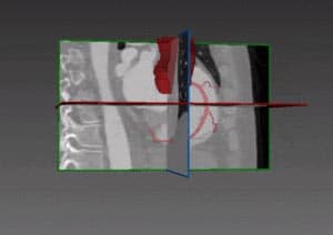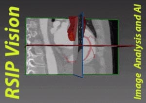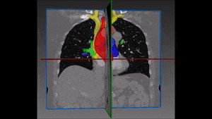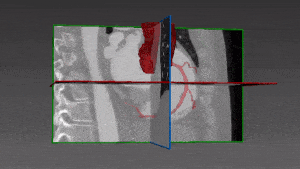
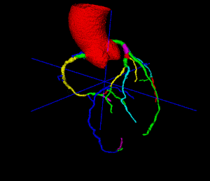

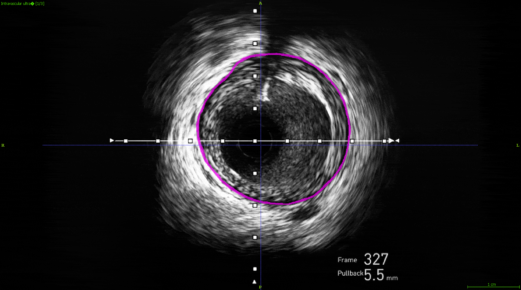
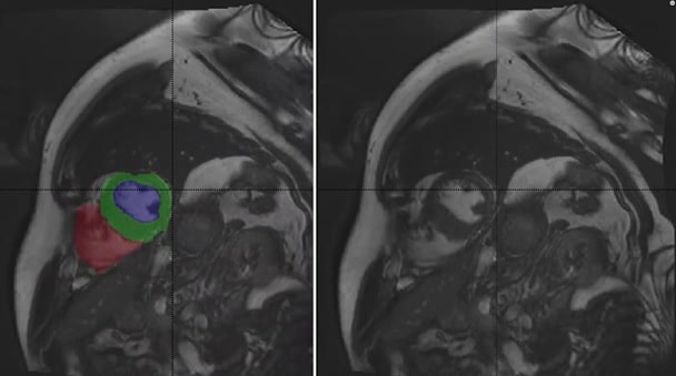
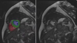

 Deep-learning based analysis
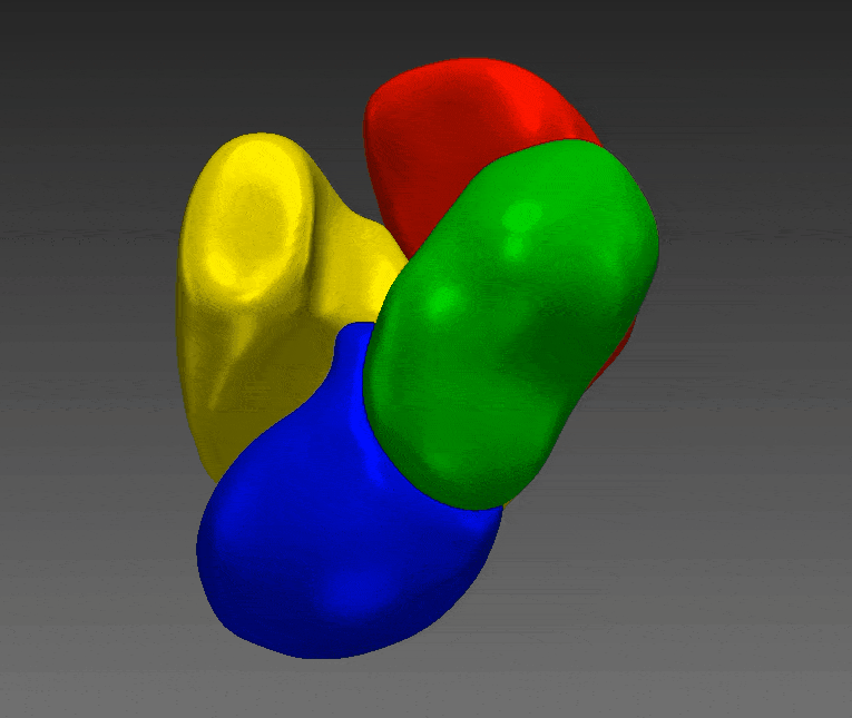
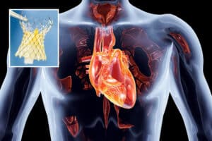
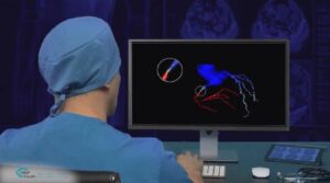
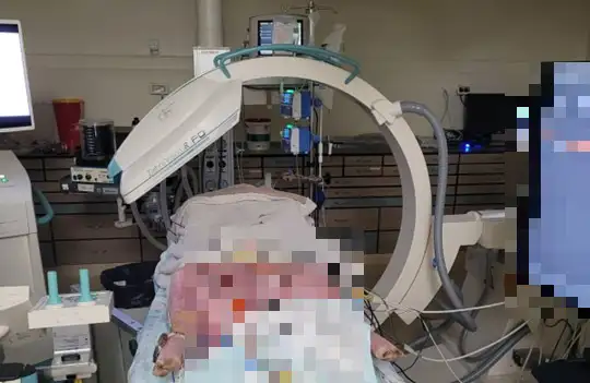
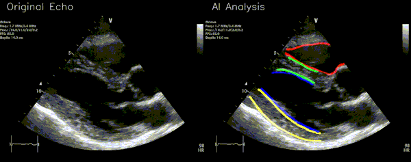
AI for Structural Heart Procedures
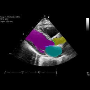
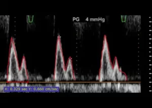
Structural heart diseases include structural deformation of the heart, like valve leakage: the blood flows in two directions and the patient is losing efficiency of the heart. These problems can lead down the line to heart failure. Cardiology offers many possible solutions to structural heart diseases, like implanting a new valve, fix the current valve, fix the valve leaflets and more. When the heart contracts, the leaflets are expected to seal adequately and direct the blood in the desired direction. When there is a malformation or problem with the leaflet,