Skip to content
  • Our Work
    • Fields
      • Cardiology
      • ENT
      • Gastro
      • Orthopedics
      • Ophthalmology
      • Pulmonology
      • Surgical
      • Urology
      • Other
    • Modalities
      • Endoscopy
      • Medical Segmentation
      • Microscopy
      • Ultrasound
  • Success Stories
  • Insights
    • Magazine
    • Upcoming Events
    • Webinars
    • Meetups
    • News
    • Blog
  • The company
    • About us
    • Careers
Menu
  • Our Work
    • Fields
      • Cardiology
      • ENT
      • Gastro
      • Orthopedics
      • Ophthalmology
      • Pulmonology
      • Surgical
      • Urology
      • Other
    • Modalities
      • Endoscopy
      • Medical Segmentation
      • Microscopy
      • Ultrasound
  • Success Stories
  • Insights
    • Magazine
    • Upcoming Events
    • Webinars
    • Meetups
    • News
    • Blog
  • The company
    • About us
    • Careers
Contact

Announcement – New Urological AI Tool for 3D Reconstruction of the Ureter

RSIP Vision Presents New Urological AI Tool for 3D Reconstruction of the Ureter Improving Urological Procedures

Innovative technology utilizes 2D fluoroscopic images and reconstructs an accurate 3D model of the ureter for planning and guidance during ureteroplasty.

TEL AVIV, Israel & SAN JOSE, Calif., October 4, 2022 – RSIP Vision, a leader in driving innovation for medical imaging through advanced AI and computer vision solutions, announces today a new tool for 3D reconstruction of the ureter. This tool transforms 2D fluoroscopic images of the ureter into a medical grade 3D model. The reconstructed model will be  used by the physician for diagnosis, procedural planning, and real-time navigation during a procedure. This new vendor-neutral technology will be available to third-party medical device manufacturers using C-arm imaging  and viewer solutions, providing an advanced 3D tool for a variety of urologic procedures.

“Creating a 3D model from 2D images is a challenging task and requires vast experience and expertise.” said Ron Soferman, CEO at RSIP Vision. “By developing advanced deep-learning (DL) algorithms we managed to reconstruct an accurate 3D model of the ureter from 2D images. The inputs to the system are at least 2  fluoroscopic images of the ureter from different acquisition angles, and the output is a CT-grade 3D model of the ureter . This can be used in a variety of procedures in urology to assist the urologist in real time.”

Ureteroplasty, in addition to other urological procedures, utilizes a retrograde or intravenous pyelogram for visualization of the ureter. Often the pyelogram is the first step of a longer procedure requiring navigation through the ureter. The images from the pyelogram are 2D fluoroscopy images, lacking the depth dimension. As shown in this video, RSIP Vision’s new tool reconstructs an accurate 3D model from the 2D images which can be used as a reference “map” by the physician. This model can be used to improve accuracy during other urological procedures as well, such as stent placement, stone localization and retrieval, or general anatomical characterization.

“There is a great anatomical variability in ureter structure between patients,” said Dr. Rabeeh Fares, a clinical fellow at the medical imaging department at St. Joseph’s Health Centre, London, Ontario, Canada. “The ability to accurately model the ureter in real-time increases the physician’s confidence during the procedure. It will allow us to safely maneuver through the ureter, even in complex anatomies, and successfully perform the desired tasks. This tool ultimately reduces complication rates and improves patient outcomes.” 

Share

Share on linkedin
Share on twitter
Share on facebook

More News

Prostate Guidance

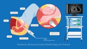
Intra-op Prostate Guidance by RSIP Vision

Automated IBD Scoring

IBD Scoring – Clario, GI Reviewers and RSIP Vision Team Up

Neph - Partial Nephrectomy surgery

RSIP Neph Announces a Revolutionary Intra-op Solution for Partial Nephrectomy Surgeries

Benign Prostatic Hyperplasia BPH

AI for Benign Prostatic Hyperplasia BPH

Knee 3D

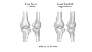
Announcement – RSIP Vision Presents Successful Preliminary Results from Clinical Study of 2D-to-3D Knee Bones Reconstruction

Stone tracking - urology

Stone tracking during kidney stone removal

Show all

RSIP Vision

Field-tested software solutions and custom R&D, to power your next medical products with innovative AI and image analysis capabilities.

Read more about us

Get in touch

Please fill the following form and our experts will be happy to reply to you soon

Recent News

IBD Scoring – Clario, GI Reviewers and RSIP Vision Team Up

RSIP Neph Announces a Revolutionary Intra-op Solution for Partial Nephrectomy Surgeries

Announcement – RSIP Vision Presents Successful Preliminary Results from Clinical Study of 2D-to-3D Knee Bones Reconstruction

Announcement – New Urological AI Tool for 3D Reconstruction of the Ureter

All news
Upcoming Events
Stay informed for our next events
Subscribe to Our Magazines

Subscribe now and receive the Computer Vision News Magazine every month to your mailbox

 
Subscribe for free
Follow us
Linkedin Twitter Facebook Youtube

contact@rsipvision.com

Terms of Use

Privacy Policy

© All rights reserved to RSIP Vision 2023

Created by Shmulik

  • Our Work
    • title-1
      • Ophthalmology
      • Uncategorized
      • Ophthalmology
      • Pulmonology
      • Cardiology
      • Orthopedics
    • Title-2
      • Orthopedics
  • Success Stories
  • Insights
  • The company