Skip to content
  • Our Work
    • Fields
      • Cardiology
      • ENT
      • Gastro
      • Orthopedics
      • Ophthalmology
      • Pulmonology
      • Surgical
      • Urology
      • Other
    • Modalities
      • Endoscopy
      • Medical Segmentation
      • Microscopy
      • Ultrasound
  • Success Stories
  • Insights
    • Magazine
    • Events and Webinars
    • News
    • Blog
  • The company
    • About us
    • Careers
Menu
  • Our Work
    • Fields
      • Cardiology
      • ENT
      • Gastro
      • Orthopedics
      • Ophthalmology
      • Pulmonology
      • Surgical
      • Urology
      • Other
    • Modalities
      • Endoscopy
      • Medical Segmentation
      • Microscopy
      • Ultrasound
  • Success Stories
  • Insights
    • Magazine
    • Events and Webinars
    • News
    • Blog
  • The company
    • About us
    • Careers
Contact

Detection and quantification of bone cancer

Primary malignant tumor of the bone (osteosarcoma) is the most common bone tumor, representing 88% of all bone tumors with nearly 3,000 annual diagnosed cases in the United States. Despite the relative low occurrence of primary osteosarcoma (less than 1% of all tumors), secondary (metastatic) bone malignancies arise following prostate cancer (80% of cases, with 3% five-year survival rate), breast cancer (with no cure) or lung cancers (with 11% two-year survival rate). Bone metastases affect more than 400,000 people annually in the United States with frequent occurrences among patients undergoing irradiation and secondary effect to other treatments.

Osteosarcoma

The detection of skeletal metastases has a major impact on devising treatment strategies and prognosis. Patients with primary or metastatic osteosarcoma are commonly cured by induction of chemotherapeutic agents and their treatment is complemented by percutaneous radiofrequency ablation. Besides the detection of primary osteosarcoma, scanning for bone metastases is prevalent for patients at high risk. Moreover, follow-up and complementary treatments require precise localization of cancerous region both near cartilage and in bone to alleviate pain caused by bone abnormalities and to locate skeletal regions at high risk of fractures.
Several scanning modalities are used to investigate and detect skeletal abnormalities. Most commonly, one finds in clinical practice low-dose CT which is used to detect deviations from normal bone structure, MRI to detect bone marrow and other soft tissues abnormalities as well as PET CT to investigate into the metabolic activity of suspected tissues. To reduce the cumulative exposure risk, clinical practice has recently been shifted toward whole-body imaging. However, resolution in such images and the low dose used render complete localization of suspected regions difficult. Precise and automated methods, able to improve the ability of an expert to pinpoint abnormalities, are thus called for.

Quantification of primary and metastatic bone cancer

With the advancement of image processing and computer vision methodologies, an alert system can be constructed by algorithmic means. Such systems are designed to automatically localize metastatic and primary cancerous regions. Quantitative characterization of the disease can then enable oncologists to better assess prognosis, which should then translate into personalized treatment, higher survival rate and improved quality of life.
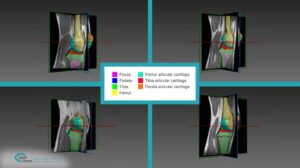
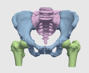
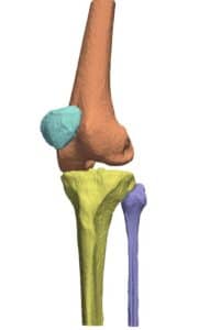
The detection procedure begins with skeletal segmentation and its distinction from surrounding anatomical parts. For this end, incorporation of knowledge into the anatomical part is useful to determine sensitivities related to bone density and relative intensity as seen by the image modality. In whole-body imaging, it is advisable to distinguish body parts and perform segmentation on each part. By itself, the recognition of body parts is not trivial. However, owing to the predicted patient pose at the time of imaging, coupled with computational models of human anatomy, such a task is indeed feasible.
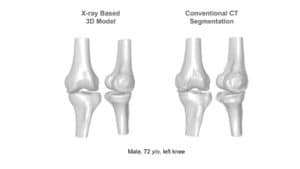
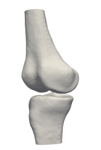
The availability of successive stacked 2D images enables iterative segmentation to produce 3D surfaces of bones and other skeletal-related structures. Stitching 2D slices accurately into a 3D model requires a primary registration and matching step to align slices and fix their relative position to other organs. Methodologies for 3D reconstruction are widely used in CAD/CAM for computer assisted surgery (CAS) and provide oncologists with a more complete image of the skeletal structure, to assist in quick detection of structural anomalies.
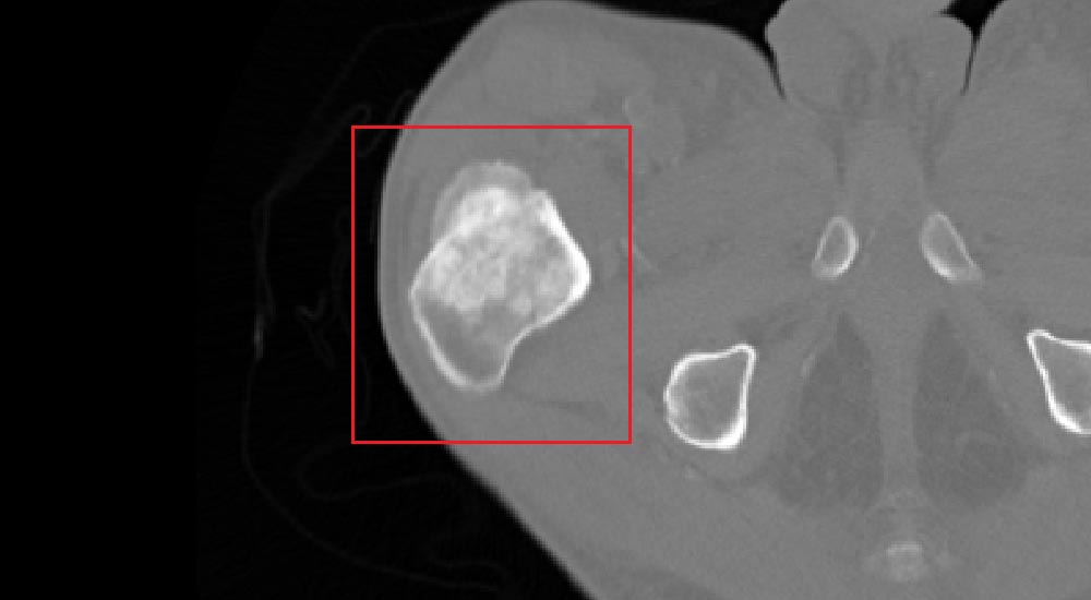
Metastatic lesions detection is performed on the segmented skeletal structure in two and three dimensions. The challenge faced by algorithms is to simultaneously extract the geometrical properties of the lesions and classify candidate structures into their severity levels. Classification of structures can be successfully performed by utilizing Support Vector Machine-based (SVM) algorithms, which ‘learn’ by way of supervision the characteristics of a set of normal and abnormal manually classified lesions. Learning process can be regarded as synonymous to adjusting weights of the SVM, once the set enables autonomous classification of newly encountered suspected structures.
The pipeline shortly outlined above by no means describes all challenges to be faced during the construction of such algorithms. In light of the accumulative information of patient history, prognostic factors and multimodal data, algorithmic means seem to be the only reasonable way to coherently incorporate all this info into the diagnosis and treatment planning. Rich of its long experience in automatic detection, 3D reconstruction and multimodal registration, RSIP Vision can help you succeed in all your algorithmic projects.

Share

Share on linkedin
Share on twitter
Share on facebook

Main Field

Orthopedics

RSIP Vision brings specific field-tested experience in solving real world orthopedic imaging challenges for multiple clinical use cases. From preoperative planning, to intraoperative navigation, and many more. We are a proven leader, enabling our clients to bring innovative AI capabilities into the market. XPlan - See our most advanced AI-enabled 3D reconstruction from standard 2D X-rays, revolutionizing joint replacement, at www.xplan.ai

View Orthopedics

Categories

  • Orthopedics, RSIP Vision Learns

Related Content

XPlan Image

Announcement – XPlan.ai Confirms Premier Precision in Peer-Reviewed Clinical Study of its 2D-to-3D Knee Reconstruction Solution

Knee 3D

Announcement – XPlan.ai by RSIP Vision Presents Successful Preliminary Results from Clinical Study of it’s XPlan 2D-to-3D Knee Bones Reconstruction

Intraoperative Registration Module for Orthopedic Surgery

Announcement – Registration Module for Orthopedic Surgery

Automated Assessment of Cartilage Damage

Announcement – Automated Assessment of Cartilage Damage

Hip

Segmentation in Orthopedics with Deep Learning

Knee

Computer-Assisted Joint Replacement (hip and knee)

Show all

RSIP Vision

Field-tested software solutions and custom R&D, to power your next medical products with innovative AI and image analysis capabilities.

Read more about us

Get in touch

Please fill the following form and our experts will be happy to reply to you soon

Recent News

Announcement – XPlan.ai Confirms Premier Precision in Peer-Reviewed Clinical Study of its 2D-to-3D Knee Reconstruction Solution

IBD Scoring – Clario, GI Reviewers and RSIP Vision Team Up

RSIP Neph Announces a Revolutionary Intra-op Solution for Partial Nephrectomy Surgeries

Announcement – XPlan.ai by RSIP Vision Presents Successful Preliminary Results from Clinical Study of it’s XPlan 2D-to-3D Knee Bones Reconstruction

All news
Upcoming Events
Stay informed for our next events
Subscribe to Our Magazines

Subscribe now and receive the Computer Vision News Magazine every month to your mailbox

 
Subscribe for free
Follow us
Linkedin Twitter Facebook Youtube

contact@rsipvision.com

Terms of Use

Privacy Policy

© All rights reserved to RSIP Vision 2023

Created by Shmulik

  • Our Work
    • title-1
      • Ophthalmology
      • Uncategorized
      • Ophthalmology
      • Pulmonology
      • Cardiology
      • Orthopedics
    • Title-2
      • Orthopedics
  • Success Stories
  • Insights
  • The company