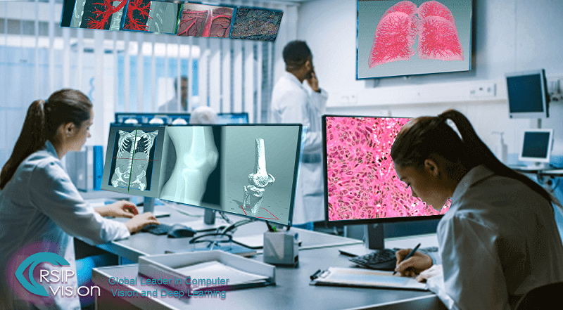
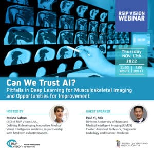
Find out how Artificial Intelligence tackles the huge diversity in the most complex challenges that exist in the medical field.
We’re happy to meet again on February 5 to discuss everything that’s new in Computer Vision, Deep Learning & Artificial Intelligence.
At this Meetup we will be discussing AI in Medical Imaging, with two great speakers (see bios below):
Agenda:
18:00-18:30 Arrival, mingling, and pizza
18:30-19:00 Dr. Sergio Sanabria – Stanford University:
Next Generation Cancer Diagnosis – Empowering Ultrasound as a Ubiquitous High-Precision Diagnostics Tool
19:00-19:30 Dr. Qingyu Zhao – Stanford University:
Confounder-aware Deep Learning in Neuroimaging Studies (see abstract below)
19:30-20:00 – Q&A and further networking
Venue: Quinlan Community Center, Cupertino.
10185 N Stelling Rd · Cupertino, CA
This meetup is made possible thanks to sponsorship by RSIP Vision. Please register below!
Abstract of Dr. Zhao’s talk: Leveraging labeled big data and enhanced computational power, deep convolutional neural networks have been applied in many neuroscience studies to advance the mechanistic understanding of brain disorders. Despite the success, one common pitfall is the negligence of algorithmic bias introduced by deep learning models towards confounding factors in the study. A confounding factor (or confounder) correlates with both the independent variable (e.g, MR image) and dependent variable (e.g. diagnosis label), thereby causing spurious association. In this talk, I will present several models that handle confounders in all stages of deep learning applications in neuroscience. Specifically, I will discuss how to learn confounder-aware generative models for image synthesis, how to train deep learning models that are unbiased towards confounders, and how to remove confounding effects from true disease effects in interpreting learned models.
Bio: Dr. Qingyu Zhao is a postdoctoral research fellow in the Department of Psychiatry and Behavioral Sciences, Stanford University. His research has been focusing on developing novel image analysis and machine learning techniques to enhance understanding, diagnosis, and treatment of neuropsychiatric disorders.
Abstract of Dr. Sanabria’s talk: At the Zurich Ultrasound Research and Translation group (www.zurt.ch), we are developing the ultrasound technology of the future. Ultrasound is a non-invasive, cost-effective imaging technology, with the potential to become ubiquitous. Hardware has already evolved into portable systems that in the near future will be running on smartphones. Yet, current ultrasound echographs are only fit for qualitative assessment of the body. Our vision is to develop ultrasound into a ubiquitous high-precision diagnostic tool. For this purpose, we are empowering ultrasound with quantitative imaging biomarkers, which until now were only available in more invasive or involved imaging modalities, such as X-ray or Magnetic Resonance Imaging.
While conventional ultrasound images consist of a grayscale map of a few kilobytes, the time signals captured by the ultrasound probes consist of several gigabytes of data, which contain a wealth of hidden information about tissue mechanics and microstructure. To convert these large datasets into meaningful information, we combine novel quantitative features optimized from tissue bio-mechanical models, for instance images of the speed of the waves travelling through the body, with self-learned features extracted with artificial algorithms. These allow us to create new quantitative imaging sequences, that can be inserted as an add-on to existing ultrasound systems.
In this presentation, I will review technological developments and results of recent clinical studies at ZURT. We will start discussing an alternative for early-stage breast cancer diagnostics without radiation or discomfort. We will continue describing a low-cost screening tool to assess muscular disease in the aging population. Finally, I will discuss our latest work in collaboration with Stanford Medicine for early diagnostics of non-alcoholic fatty liver disease, a common yet asymptomatic cause for cirrhosis and liver cancer.
Bio: Sergio J. Sanabria received the M.Sc. degree in Telecommunications Engineering from the University of the Basque Country, Bilbao, Spain, in 2007, and the Ph.D. degree from ETH Zurich, Switzerland, in 2012. From 2012 to 2014, he was a postdoc at the Institute for Building Materials of ETH, where he developed acoustic holography methods for non-destructive testing of wood constructions materials. From 2014 to 2017 he was Senior Assistant at the Computer Vision Laboratory of ETH, where he co-led the ultrasound elastography research. Since 2018, he is pursuing his habilitation as Assistant Professor at the University Hospital of Zurich, and since October 2019, he is a visiting scholar at the Department of Radiology at Stanford School of Medicine. He has authored or co-authored over 30 peer-reviewed papers and has four patents. Dr. Sanabria received the Best Young Academics Dissertation Award from the German Society of Non-Destructive Testing (DGZfP) and the Gold Medal Award from the International Academy of Wood Science. In 2016, he received the Spark Award for the most promising invention registered for a patent at ETH Zurich for the speed-of-sound imaging technology.

